Most hip replacements are performed for osteoarthritis — the progressive loss of the cartilage covering the femoral head and the socket. It causes groin pain that can radiate to the thigh and knee, morning stiffness, and growing difficulty walking, putting on shoes and climbing stairs. It does not resolve on its own: conservative treatment controls symptoms, while replacement remains the definitive solution once pain limits daily life.
Other conditions I treat
- Avascular necrosis of the femoral head — the death of bone tissue caused by interrupted blood supply, which can lead to collapse of the head and secondary arthritis. Early diagnosis by MRI matters, because in the initial stages there are options that may avoid replacement.
- Developmental dysplasia of the hip — a socket too shallow to contain the femoral head properly, concentrating load on a reduced surface and causing early arthritis. In adults, replacement requires careful planning because the altered anatomy changes where the components must sit.
- Periprosthetic fracture and implant failure — see revision surgery below.
The direct anterior approach
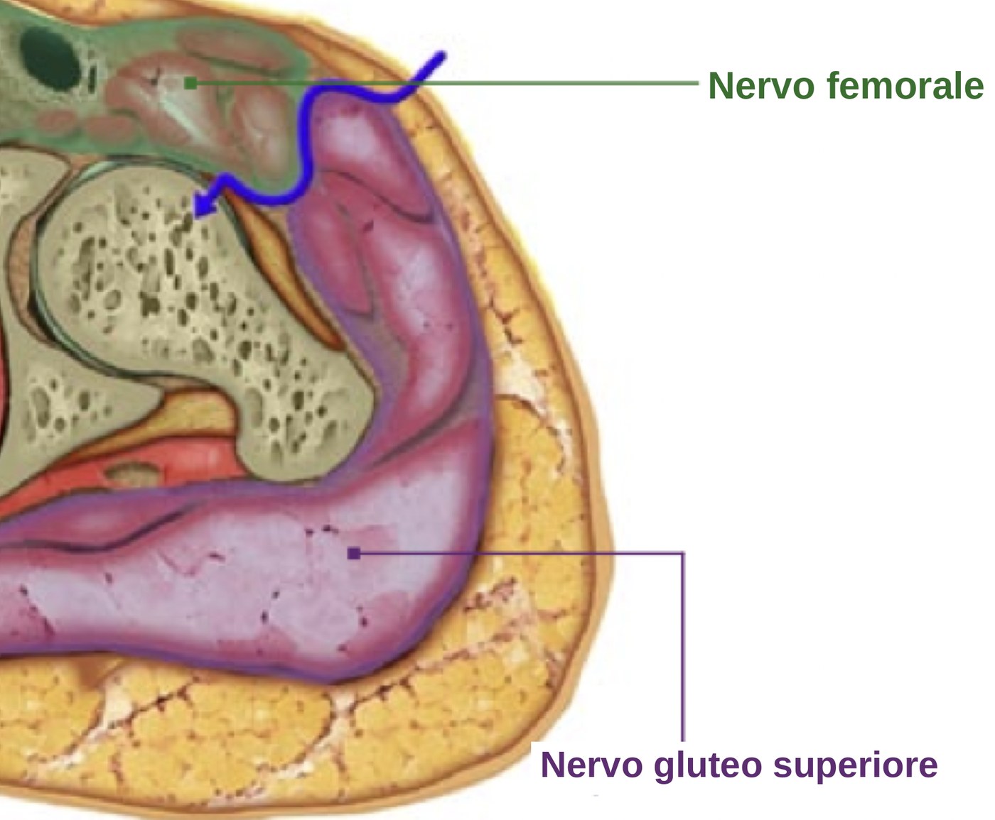
Surgical techniqueThe direct anterior approach reaches the hip by passing between muscles rather than cutting through them. It follows the Hueter interval, a genuine internervous plane: laterally the tensor fasciae latae, supplied by the superior gluteal nerve; medially the sartorius and rectus femoris, supplied by the femoral nerve. Between two different nerve territories there is always a natural boundary, and this approach uses it. No muscle is divided or detached — they are simply retracted apart.
The patient lies supine rather than on their side. That keeps the pelvis in a stable, neutral position, which makes it easier to control cup orientation and leg length, allows intraoperative fluoroscopy, and lets the two legs be compared directly.
| Compared with the posterior approach | Finding | What it means |
|---|---|---|
| Pain in the first 72 hours | in favour | Consistently lower pain scores over the first three days. |
| Function at 2–6 weeks | in favour | Better functional scores in the early weeks — the most consistent advantage. |
| Length of stay | in favour | Shorter hospital stay on average. |
| Function at 3 months and beyond | no difference | At three months, six months and one year the two groups are equivalent. The advantage is in the speed of recovery, not the final result. |
| Dislocation risk | not demonstrated | The anatomical rationale is strong — the posterior structures are left intact — but meta-analyses of randomised trials have not confirmed a statistically significant reduction. |
| Operating time | against | Longer on average, particularly during the learning curve. |
| Thigh numbness | against | A complication specific to this approach, involving the lateral femoral cutaneous nerve. Purely sensory: it never affects strength or walking. |
The approach is not right for everyone. Significant obesity, severe deformity, previous hip surgery, poor bone quality or the need for wide exposure all point towards a different route. The approach is a means to a result, not a goal in itself.
Femur First and revision
Further techniquesFemur First
A planning philosophy that reverses the usual order: the femoral component is positioned first, and the socket is then oriented to match it. Because the combined orientation of the two components determines how far the hip can move before impingement, starting from the femur — whose anatomy cannot be altered — allows the cup to be adapted to the individual rather than placed at a standard angle.
Revision hip replacement
Revision surgery replaces an implant, wholly or in part, once it has stopped working. The main causes are loosening of the components, wear of the bearing surfaces, recurrent dislocation, periprosthetic fracture and infection. It is more demanding than primary surgery: it needs dedicated planning, specific implants, and — in infection — a staged protocol.
Periprosthetic infection is the hardest scenario. Treatment usually involves two stages: removal of the implant with placement of an antibiotic-loaded spacer, a course of targeted antibiotic therapy, and reimplantation once the infection is controlled.